Home
/ Back Muscles Anatomy Chart - Muscle Anatomy Skeletal Muscles Human Anatomy Anatomy Poster Skeleton Human Skeleton : Low back muscle spasming is common because lumbar extensor muscles must contract eccentrically, isometrically, and concentrically whenever we bend forward…
Back Muscles Anatomy Chart - Muscle Anatomy Skeletal Muscles Human Anatomy Anatomy Poster Skeleton Human Skeleton : Low back muscle spasming is common because lumbar extensor muscles must contract eccentrically, isometrically, and concentrically whenever we bend forward…
Back Muscles Anatomy Chart - Muscle Anatomy Skeletal Muscles Human Anatomy Anatomy Poster Skeleton Human Skeleton : Low back muscle spasming is common because lumbar extensor muscles must contract eccentrically, isometrically, and concentrically whenever we bend forward…. Intermediate back muscles and c. In this section, learn more about the muscles of the. The muscles of the back are a group of strong, paired muscles that lie on the posterior aspect of the trunk. Learn about these muscles, their locations there are several individual muscles within the back anatomy, and it's important to take a quick look at all of them to see how you can target them. To build the back optimally, you should know the major muscles, their actions, and which exercises build muscles best.
William is a final year medical student in australia who has taught anatomy to tertiary science and medical students since 2010. Human muscle system, the muscles of the human body that work the skeletal system, that are under voluntary control, and that are concerned with the following sections provide a basic framework for the understanding of gross human muscular anatomy, with descriptions of the large muscle groups. The muscles of the back are a group of strong, paired muscles that lie on the posterior aspect of the trunk. This is a table of skeletal muscles of the human anatomy. Attached to the bones of the skeletal system are about 700 named.
Best Back Exercises from www.makeoverfitness.com Attached to the bones of the skeletal system are about 700 named. Anatomy posters and anatomy charts. Facebook twitter whatsapp pin it. The superficial back muscles are the muscles found just under the skin. Scientific studies using sophisticated tools such as electromyography (emg) and. Muscles, connected to bones or internal organs and blood vessels, are in charge for movement. Muscle charts female muscle mini. Microscopic anatomy of skeletal muscle.
The deep back muscles lie immediately adjacent to the vertebral column and ribs.
Musculature anatomy chart poster laminated. Let's remember the back muscles. Their main function is contractibility. Select a region pectoral superficial back & scapular arm anterior forearm posterior forearm hand. Other sets by this creator. They provide movements of the spine functional anatomy: While working on neck muscles. The muscular system is responsible for the movement of the human body. Intermediate back muscles and c. They are divided into three groups, as shown below. This anatomy chart is a great example of beauty and function in one, as it is pleasing to look at and is very educational. This entry was posted in anatomy, muscles and tagged anatomy of back muscles, back muscles anatomy, back muscles chart, back muscles diagram, back muscles diagram with labels, back muscles explained, human. Muscle charts female muscle mini.
Intermediate back muscles and c. Front view of muscles, skeleton, organs, nervous system. Anatomy posters and anatomy charts. Learn about these muscles, their locations there are several individual muscles within the back anatomy, and it's important to take a quick look at all of them to see how you can target them. The deep back muscles lie immediately adjacent to the vertebral column and ribs.
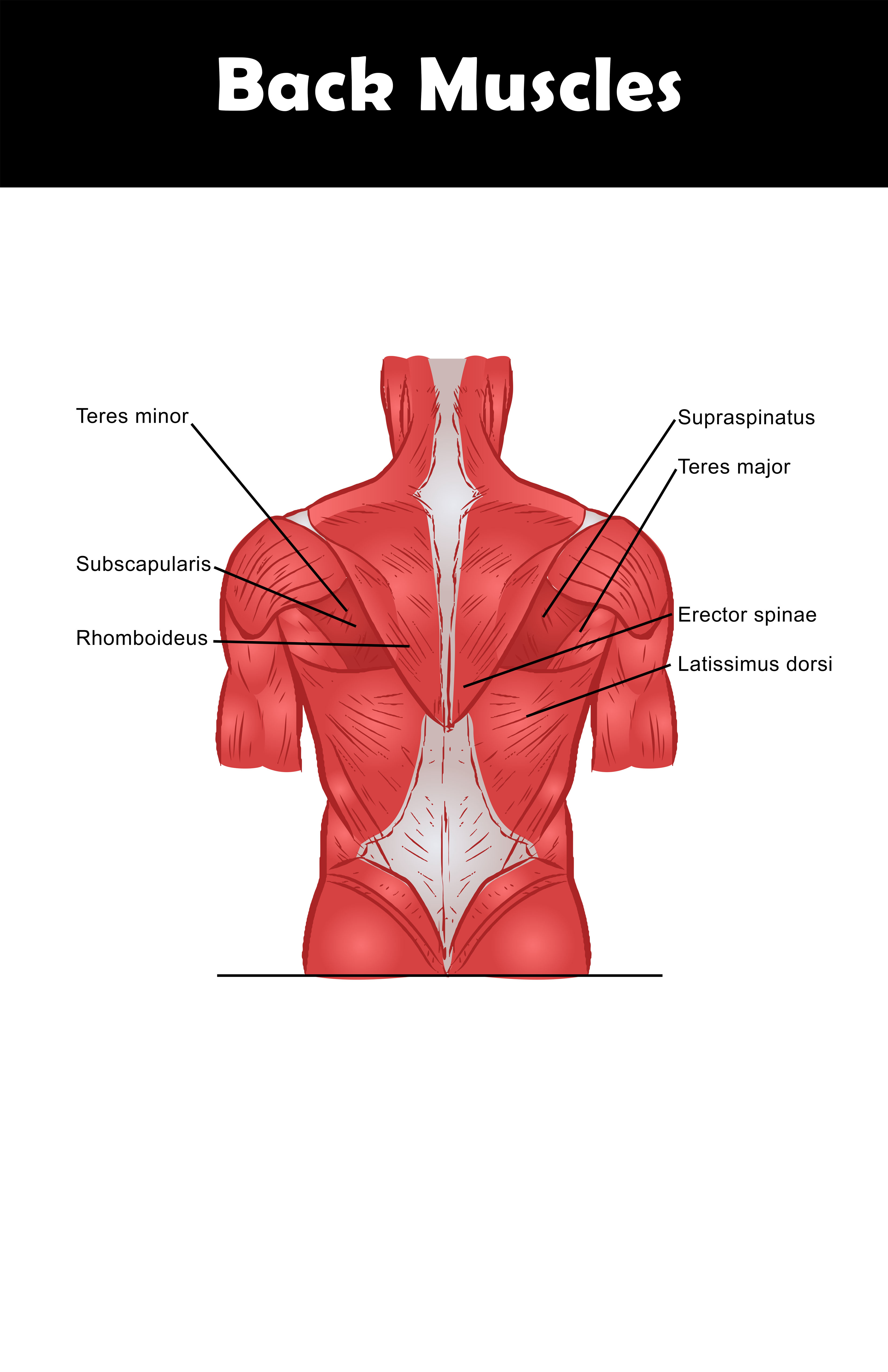
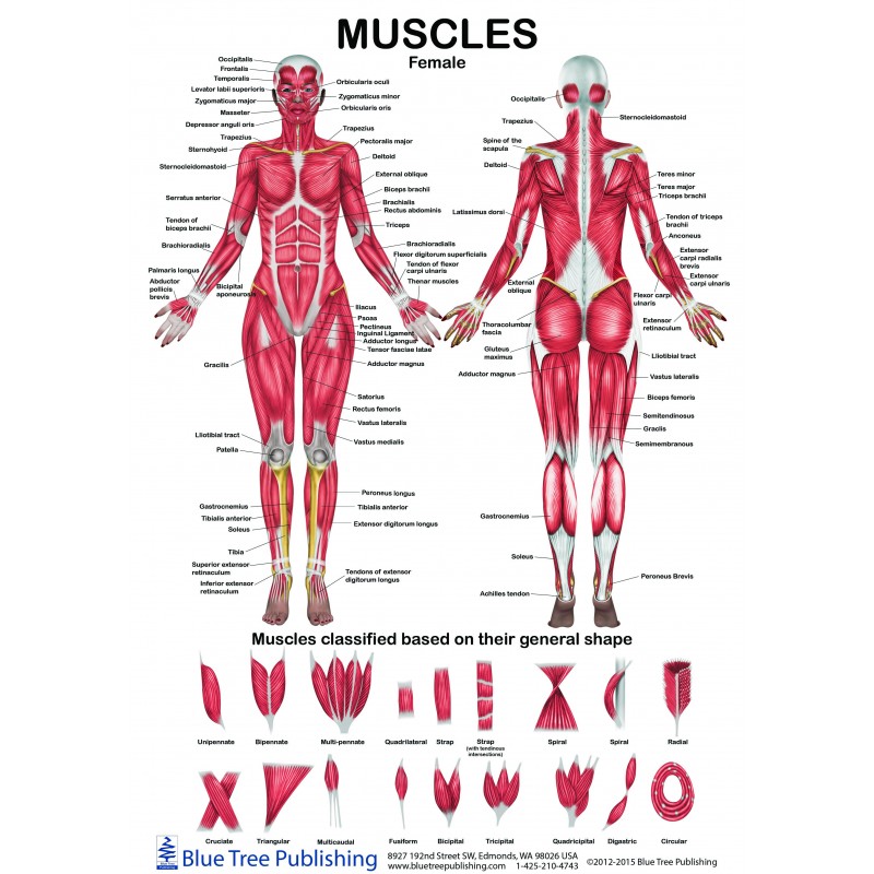
Female Male Muscle Anatomical Chart from www.bluetreepublishing.com Anatomical diagram showing a back view of muscles in the human body. Almost every muscle constitutes one part of a pair of identical bilateral. They provide movements of the spine functional anatomy: This entry was posted in anatomy, muscles and tagged anatomy of back muscles, back muscles anatomy, back muscles chart, back muscles diagram, back muscles diagram with labels, back muscles explained, human. Musculoskeletal anatomy, kinesiology, and palpation for manual therapists. To build the back optimally, you should know the major muscles, their actions, and which exercises build muscles best. Labeled anatomy chart of neck and back muscles on white. Low back muscle spasming is common because lumbar extensor muscles must contract eccentrically, isometrically, and concentrically whenever we bend forward…
The teres major muscle originates on the outer (lateral) edge of the scapula and attaches to the humerus.
This is a table of skeletal muscles of the human anatomy. Muscles of the back, anatomy chart. The muscles of the back that work together to support the spine, help keep the body upright and allow twist and bend in many directions. The teres majo r muscles work with the rotator cuff muscles to stabilize the shoulder joint and works with the latissimus dorsi muscles to pull the humerus back. Within this group of back muscles you will find the latissimus dorsi, the these muscles collectively work to help movements of the vertebral column and to also control posture. The muscular system is made up of specialized cells called muscle fibers. Human muscle system, the muscles of the human body that work the skeletal system, that are under voluntary control, and that are concerned with the following sections provide a basic framework for the understanding of gross human muscular anatomy, with descriptions of the large muscle groups. Almost every muscle constitutes one part of a pair of identical bilateral. Here the extrinsic back muscles are classified into logical subgroups to facilitate knowledge. Labeled anatomy chart of neck and back muscles on white. Anatomy of the muscular system. Front view of muscles, skeleton, organs, nervous system. Great round muscle (tere major, tma) little round muscle (teres minor, tmi) infraspinatus muscle (is) these are the main muscle that is responsible for turning and rotating the arm.
The muscles of the back that work together to support the spine, help keep the body upright and allow twist and bend in many directions. Microscopic anatomy of skeletal muscle. The deep back muscles lie immediately adjacent to the vertebral column and ribs. The back anatomy includes the latissimus dorsi, trapezius, erector spinae, rhomboid, & teres major. This entry was posted in anatomy, muscles and tagged anatomy of back muscles, back muscles anatomy, back muscles chart, back muscles diagram, back muscles diagram with labels, back muscles explained, human.
Muscles Of The Lumbar Spine Of The Trunk from www.learnmuscles.com They are located deep to the extrinsic muscles, being. Other sets by this creator. Be sure to visit the guide for more context and information about back muscles anatomy chart, or read some of our other health & anatomy posts! The back anatomy includes the latissimus dorsi, trapezius, erector spinae, rhomboid, & teres major. While working on neck muscles. This is a table of skeletal muscles of the human anatomy. The muscles of the back that work together to support the spine, help keep the body upright and allow twist and bend in many directions. Great round muscle (tere major, tma) little round muscle (teres minor, tmi) infraspinatus muscle (is) these are the main muscle that is responsible for turning and rotating the arm.
Facebook twitter whatsapp pin it.
Select a region pectoral superficial back & scapular arm anterior forearm posterior forearm hand. William is a final year medical student in australia who has taught anatomy to tertiary science and medical students since 2010. They provide movements of the spine functional anatomy: Musculature anatomy chart poster laminated. Facebook twitter whatsapp pin it. Muscles of the back can be divided into superficial, intermediate, and deep group.since the all the back muscles originate in embryo (fetus) form by locations other than the back, muscles in the. The teres majo r muscles work with the rotator cuff muscles to stabilize the shoulder joint and works with the latissimus dorsi muscles to pull the humerus back. Scientific studies using sophisticated tools such as electromyography (emg) and. Anatomy chart courtesy of fcit. Included are several layered views of the back muscles, the doral muscles, subclavius muscles, rhomboideus major and minor muscles, deltoid muscles and many more. The teres major muscle originates on the outer (lateral) edge of the scapula and attaches to the humerus. The muscles of the back are a group of strong, paired muscles that lie on the posterior aspect of the trunk. Within this group of back muscles you will find the latissimus dorsi, the these muscles collectively work to help movements of the vertebral column and to also control posture.
Let's remember the back muscles back muscles anatomy. Front view of muscles, skeleton, organs, nervous system.
